PATIENT EDUCATION & ARTICLES•COMMUNITY HEALTH SERIES•UPDATED 2026
DOCTOR VERIFIED
KANPUR / NEW DELHI
✦HEALTH GUIDES & NEUROSURGERY ARTICLES
Understanding Brain, Spine & Nerve Health
Evidence-based clinical insights on brain, spine and nerve health to help patients and families better understand neurosurgical conditions and treatment options.
✓Written & clinically reviewed by Dr. Prashank Goel (MCh Neurosurgery, GIPMER)
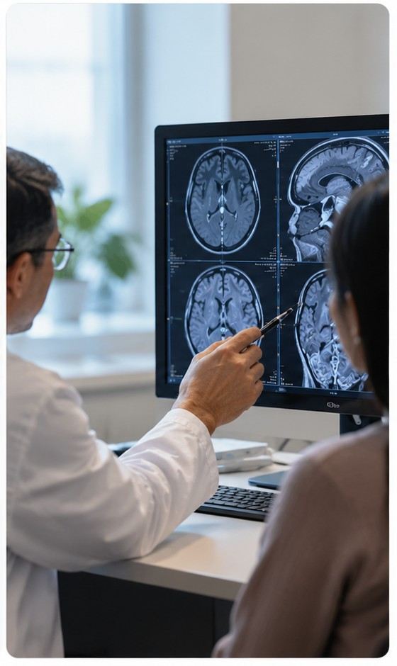
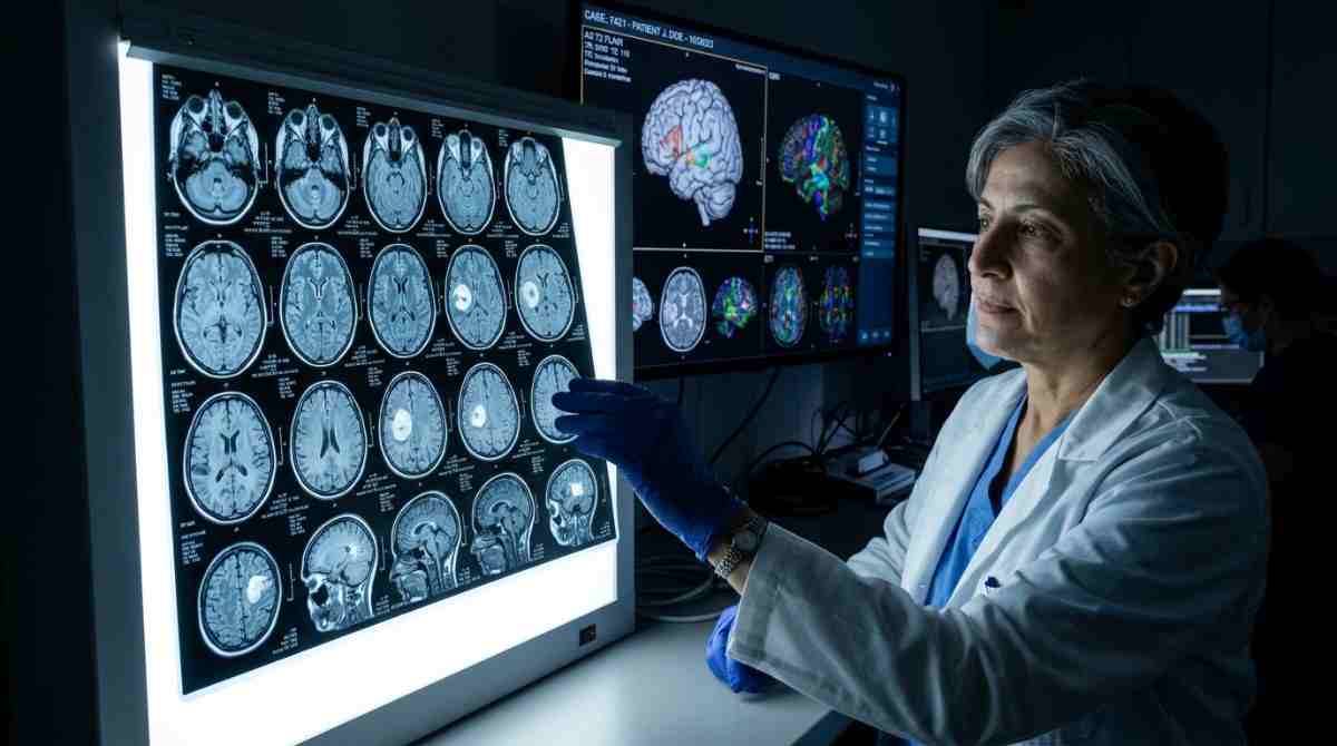
CONSULTATION & SCAN REVIEWKANPUR CLINIC
Evidence-based care meets clear, compassionate communication.Paras Health
Authored & clinically reviewed by Dr. Prashank Goel.
Minimally Invasive Spine • CLINICAL GUIDE
5 Min Read
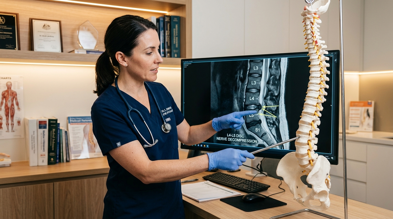
Evolution of Tubular Retractors & Minimally Invasive Spine Decompression
Academic presentation detailing microscopic tubular portals for lumbar discectomy, eliminating back muscle cutting and enabling next-day patient ambulation.
CLINICAL METHODOLOGY & APPROACH:
Longitudinal follow-up of 120 patients undergoing endoscopic and microscopic tubular decompression for far-lateral and intracanalicular lumbar disc herniations. Outcomes measured via Oswestry Disability Index (ODI) and VAS pain scores.
Academic presentation detailing microscopic tubular portals for lumbar discectomy, eliminating back muscle cutting and enabling next-day patient ambulation.
Longitudinal follow-up of 120 patients undergoing endoscopic and microscopic tubular decompression for far-lateral and intracanalicular lumbar disc herniations. Outcomes measured via Oswestry Disability Index (ODI) and VAS pain scores.
Tubular access systems preserve dorsal ramus innervation and native spinal biomechanics, demonstrating superior long-term functional recovery, minimal blood loss, and zero adjacent segment instability.
The evolution of minimally invasive spine surgery (MISS) has redefined patient recovery trajectories for lumbar canal stenosis and sciatica. By replacing large open incisions with serial muscle-dilating tubular retractors, the integrity of paraspinal musculature and stabilizing ligamentous complexes is strictly maintained.
Patients achieve dramatic reduction in post-operative wound discomfort, experience negligible blood loss, and are routinely discharged within 24 hours, returning to active life far sooner than open surgery counterparts.
CLINICAL DOSSIER ARCHIVE
Clinical Insights & Surgical Guides
Patient-friendly articles covering neurosurgical conditions, treatment options, and recovery.
Minimally Invasive Spine5 Min Read
Evolution of Tubular Retractors & Minimally Invasive Spine Decompression
Academic presentation detailing microscopic tubular portals for lumbar discectomy, eliminating back muscle cutting and enabling next-day patient ambulation.
Published: Sep 30, 2026Clinically Verified
Academic presentation detailing microscopic tubular portals for lumbar discectomy, eliminating back muscle cutting and enabling next-day patient ambulation.
Longitudinal follow-up of 120 patients undergoing endoscopic and microscopic tubular decompression for far-lateral and intracanalicular lumbar disc herniations. Outcomes measured via Oswestry Disability Index (ODI) and VAS pain scores.
Tubular access systems preserve dorsal ramus innervation and native spinal biomechanics, demonstrating superior long-term functional recovery, minimal blood loss, and zero adjacent segment instability.
The evolution of minimally invasive spine surgery (MISS) has redefined patient recovery trajectories for lumbar canal stenosis and sciatica. By replacing large open incisions with serial muscle-dilating tubular retractors, the integrity of paraspinal musculature and stabilizing ligamentous complexes is strictly maintained.
Patients achieve dramatic reduction in post-operative wound discomfort, experience negligible blood loss, and are routinely discharged within 24 hours, returning to active life far sooner than open surgery counterparts.
Comprehensive clinical series reporting anatomical variations in complex cranial bases and operative strategies to spare essential cranial nerves during intricate tumor resections.
Published: Sep 30, 2026Clinically Verified
Comprehensive clinical series reporting anatomical variations in complex cranial bases and operative strategies to spare essential cranial nerves during intricate tumor resections.
Retrospective multi-centre anatomical and surgical cohort examining complex petroclival, jugular foramen, and anterior cranial fossa surgical corridors with continuous intraoperative neuromonitoring (IONM) across cranial nerves III through XII.
Preservation of lower cranial nerve function and complete tumor extirpation are significantly enhanced through meticulous skull-base corridors, customized bone drilling, and staged resections when indicated.
Skull base surgery requires unprecedented anatomical precision to navigate around cranial nerves and major cerebral arteries. Modern approaches leverage high-definition operating microscopes, intraoperative cranial nerve monitoring, and targeted skull base drilling to achieve complete lesion resection while safeguarding neurological function.
This monograph details surgical strategies in petroclival meningiomas, acoustic neuromas, and complex vascular malformations located in deep cranial corridors.
Neurovascular Surgery7 Min Read
Intraprocedural Rupture of Cerebral Aneurysms: Effect of Anatomical Location on Surgical Outcomes
Clinical study examining intracranial aneurysm bifurcation anatomy, hemodynamic stress factors, and urgent micro-clipping rescue protocols to safeguard neurological recovery.
Published: Sep 29, 2026Clinically Verified
Clinical study examining intracranial aneurysm bifurcation anatomy, hemodynamic stress factors, and urgent micro-clipping rescue protocols to safeguard neurological recovery.
Retrospective clinical analysis of intraprocedural aneurysm ruptures treated across tertiary neurosurgical referral centres. Hemodynamic parameters, temporary parent-vessel occlusion durations, and emergency microsurgical clipping maneuvers were systematically audited against Glasgow Outcome Scale (GOS) metrics.
Intraprocedural aneurysm rupture during endovascular or micro-neurosurgical procedures represents one of the most critical neurosurgical emergencies. Understanding high-risk anatomical configurations and establishing rapid proximal vascular control safeguards brain tissue from devastating ischemia and hemorrhage.
Immediate microsurgical rescue clipping and temporary parent vessel occlusion protocols ensure optimal neurological recovery even in complex anterior communicating artery ruptures.
Spine Surgery & Diagnostics5 Min Read
Micro-Endoscopic vs. Open Spine Decompression: Indications, Biomechanics & Functional Recovery
A comprehensive guide on micro-endoscopic discectomy for lumbar disc herniations, comparing muscle-sparing tubular corridors with conventional open laminectomy.
Published: Sep 29, 2026Clinically Verified
A comprehensive guide on micro-endoscopic discectomy for lumbar disc herniations, comparing muscle-sparing tubular corridors with conventional open laminectomy.
Comparative clinical series evaluating patients undergoing targeted tubular micro-discectomy (16-18mm tubular corridor) versus open laminectomy. Clinical endpoints evaluated operative blood loss, post-operative analgesic requirements, length of hospital stay, and duration until unrestricted ambulation.
Tubular micro-endoscopic decompression achieved equivalent neural decompression with an 85% reduction in surgical blood loss and shortened median hospitalization to 24-36 hours. Sparing the multifidus musculature and posterior ligamentous complex significantly diminished long-term back stiffness and eliminated adjacent-segment instability.
Advancements in uniportal and biportal spinal endoscopy have revolutionized the management of lumbar disc herniation and spinal canal stenosis. Patients experience significantly reduced blood loss, preservation of posterior paraspinal musculature, and rapid mobilization within 24 to 48 hours post-procedure.
By utilizing targeted 16-18mm tubular retractor corridors, surgeons decompress impinged thecal sacs and exiting nerve roots under direct high-magnification visualization without destablizing posterior bony structures.
Brain Surgery & Neuro-Oncology6 Min Read
Role of Retinal Nerve Fibre Layer (RNFL) in Post-Operative Visual Recovery in Pituitary Adenomas
Optical Coherence Tomography (OCT) study correlating pre-operative peripapillary RNFL thickness with the rate of visual field restoration following endoscopic transsphenoidal adenoma resection.
Published: Sep 29, 2026Clinically Verified
Optical Coherence Tomography (OCT) study correlating pre-operative peripapillary RNFL thickness with the rate of visual field restoration following endoscopic transsphenoidal adenoma resection.
Prospective clinical study evaluating patients undergoing microscopic and endoscopic transsphenoidal decompression. Pre-operative and serial post-operative RNFL thicknesses across superior, inferior, nasal, and temporal quadrants were analyzed in correlation with visual evoked potentials (VEP) and automated Humphrey visual field recovery at 1, 3, and 6-month surgical milestones.
Patients with preserved pre-operative mean RNFL thickness achieved substantial post-operative visual field and acuity recovery. In contrast, severe pre-operative axonal thinning indicated irreversible optic nerve atrophy, confirming that timely endoscopic transnasal decompression before structural axonal loss is essential to restore sight.
Pituitary adenomas frequently compress the optic chiasm, causing insidious visual field defects (bitemporal hemianopia) and progressive loss of visual acuity. While automated perimetry maps visual defects, Optical Coherence Tomography (OCT) measurement of peripapillary Retinal Nerve Fibre Layer (RNFL) thickness provides an objective, quantitative biomarker to forecast long-term visual restoration after endoscopic transsphenoidal resection.
Patients with preserved pre-operative RNFL thickness demonstrated superior post-operative visual recovery, confirming that early surgical decompression before axonal atrophy is crucial for optimal visual outcomes.
✦INDEX OF SCHOLARSHIP // PEER-REVIEWED ARCHIVE
Research & Academic Contributions
A selected record of research, academic presentations, and neurosurgical contributions.
All Contributions (2)Brain & PituitaryNeurovascular & AneurysmsSpine & Decompression
ACADEMIC INVESTIGATORREGISTRATION NO. 63003 (UP Medical Council / MCI)
Dr. Prashank Goel
Consultant Neurosurgeon
Neurosurgical practice with academic and research contributions across brain, spine, and neurovascular care.
Paras Health & Swaroop NagarKanpur, India
14+
Surgical Exp
2x
Conclave Awards
Presenter
Paper & Poster
Superspecialty Pedagogy:GIPMER (MAMC), Delhi
Advanced Specialization:Endoscopic & Vascular
Institutional Role:Associate Director
★ 2nd Best Paper Award • 2019-02-01
Neurosurgery & Neuro-Ophthalmology
Role of Retinal Nerve Fibre Layer Thickness in Assessing Post-Operative Visual Recovery in Pituitary Adenomas
Received the 2nd Best Paper Award for research examining the role of retinal nerve fibre layer thickness in assessing post-operative visual recovery in patients with pituitary adenomas.
21st Annual Conference of Delhi Neurological Association (DNA)
★ 2nd Best Poster Award • 2019-02-01
Neurosurgery & Cerebrovascular Surgery
Intraprocedural Rupture of Cerebral Aneurysms: Effect of Location on Outcome
Received the 2nd Best Poster Award for research on intraprocedural rupture of cerebral aneurysms and the relationship between aneurysm location and clinical outcomes.
Cerebral Aneurysm Rupture & Clinical Outcomes
21st Annual Conference of Delhi Neurological Association (DNA)
DIAGNOSTIC SECOND OPINION & EVIDENCE EVALUATION
Have an MRI or CT Scan Needing Specialist Evaluation?
Connect with Dr. Prashank Goel for specialist review of your scans and discussion of appropriate treatment options.